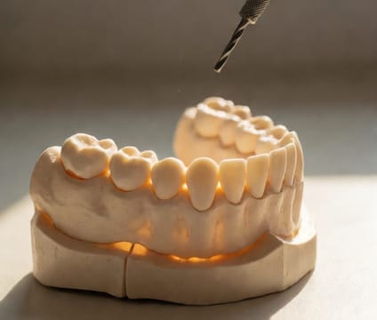
Tap to See: Osteotomy Depth & Angulation
Proper drilling angle and depth prevent sinus wall perforation and ensure implant placement safety in the upper back jaw.
Read more →This interactive 3D walkthrough presents a real-anatomy, CBCT-based visualization of upper jaw implant placement. Drag to rotate, tap to advance steps, and toggle overlays to see bone layers, nerves, and sinus relationships. Every action reflects clinical decision-making—like how angulation prevents sinus breach or why drilling depth varies by bone quality. It’s designed to clarify what happens before, during, and after the implant is placed, so you walk into your consultation with clear, grounded expectations.
Proper drilling angle and depth prevent sinus wall perforation and ensure implant placement safety in the upper back jaw.
Read more →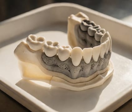
Bone quality (D1–D4) directly impacts how firmly an implant holds at placement and how much torque is safely applied.
Read more →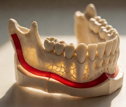
The mandibular canal migrates upward and forward with aging; our model adapts to patient age for safer lower implant planning.
Read more →Too many patients proceed with implants after one opinion—often without reviewing their CBCT scans or considering alternatives like bridge vs. implant vs.…