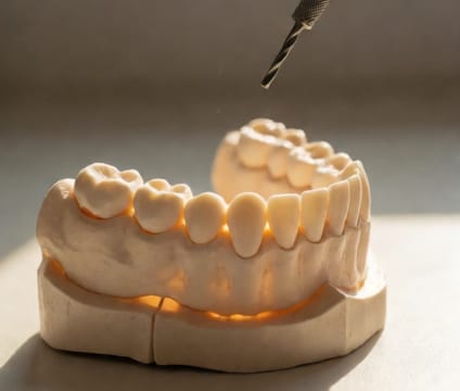
Tap for: Nerve Avoidance Zones
The mandibular canal migrates upward and forward with aging; our model adapts to patient age for safer lower implant planning.
In younger patients, the mandibular canal runs close to the root apices of lower molars. With age, bone resorption and tooth loss cause the canal to shift upward toward the mandibular ridge—sometimes placing it only 2–3 mm beneath the alveolar crest. numbness or neuropathic pain. Our interactive feature lets you select age ranges (35, 55, 75) to see how canal height and curvature shift across generations.
Implant planning must account for the canal's changing position. At age 35, a 10 mm implant might clear the canal safely; at 75, the same length could invade it due to vertical ridge loss. We highlight safe insertion depths and angulation limits for each age group, using CBCT-derived canal mapping to simulate the true 3D route. Tapping on the canal reveals its distance from the planned osteotomy site at each step.
Avoiding nerve injury isn’t just about length—it’s about path. A vertical drill trajectory is riskier than a slightly palatal one if the canal is superiorized. Our model overlays the neurovascular bundle in translucent red, showing how minor angulation corrections keep the implant 2–4 mm clear. Patients who see this in 3D better understand why a quick scan beats a generic protocol.